Scientific Program

Conference Series Ltd invites all the participants across the globe to attend 18th International Conference on Oral Health and Maxillofacial Surgery Madrid, Spain.

Day 1 :

Keynote Forum

Ahmed Halim Ayoub

Egyptian society of oral Implantology, Egypt

Keynote: 4D Concept and Immediate Implant Placement
Conference Series Oral Surgery  2016 International Conference Keynote Speaker Ahmed Halim Ayoub photo
Biography:

Ahmed Halim Ayoub. DDS. MSc. Visiting Lecturer in BPP University Faculty of Dentistry, London, England. His Diploma and Master Degrees are from Seville university, Spain . He developed expertise in bone augmentation and sinus elevation. He has a special interest in bone healing, role of growth factors and bone morphogenetic proteins in bone growth. Dr Halim is also highly interested in Dental implant Education and developed his pioneer modules using Live training throughout directing dental implant post graduate training in Egyptian society of oral Implantology, London oral restorative academy ,and London dental education services. Prof Halim is a distinguished speaker; he presents in international conferences and teaches in the UK, Europe and the Middle East.
 

Abstract:

The 4th additional axis of time is added to the traditional 3D axes which makes it different to identify the time of extraction and implant placement either immediate implant placement , early implant placement or delayed implant placement. This review focus on each technique and the ideal choice of each technique in every case
Introduction: Immediate implant placement after extraction has become a favored treatment protocol with many clinicians worldwide. There are many advantages to this protocol, amongst them; shortened treatment time, placement of the implant in sound bone that constitutes the socket wall, placement trajectory guidance by the socket and preservation of bone volume. This literature review describes the 4th dimension in implant placement which is the timing of placement after extraction which is a very important factor in immediate placement success rate.
Learning objectives:
1. After the presentation the audience shall understand:
2. The 4th dimension in implant placement
3. The proper way achieve an perfect immediate implant
4. Decide whether to place or not to place the implant after extraction
5. Manage the jumping gap
6. The proper way to achieve the maximum esthetic outcome in immediate placement.

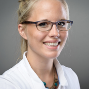
Conference Series Oral Surgery  2016 International Conference Keynote Speaker Philine H Metelmann photo
Biography:

Philine H Metelmann earned her Degree in Dentistry at Greifswald University, Germany. She completed her Doctorate in the field of Comprehensive Cancer Prevention for which she received the nomination for Felix Burda Award 2015. In 2012 she was awarded a grant from DAAD (German Academic Exchange Service) to continue her research and education as a visiting student at University of Minnesota, USA. After working in a private practice she is currently doing an Orthodontic Residency and carries out different research projects.

Abstract:

This retrospective study describes the craniofacial architecture of post-pubertal patients with cleft lip and palate by using DELAIRE whole skull analysis along with the analysis by SEGNER AND HASUND. 177 cephalometric X-Ray films from a group of patients (114 male, 63 female) of the former Wolfgang Rosenthal Clinic Thallwitz, Germany, were analyzed. Typical changes in the viscero-cranium (mandibular and maxillary retrognathia in a vertical basal open relationship) were accompanied by small yet statistically significant changes to the neuro-cranium. The study revealed mid-face deficits in the sagittal plane and a reduced height of the mid-face in favour of the lower face. Furthermore, a reduced craniofacial base line and cranial height and a reduced angle between the cranial base and craniofacial base line were evident. The angle between the cranial base and the basilar slope was enlarged, suggesting an abnormal posture of the cervical spine in CLP patients. Cleft lip and palate along with late-closure corrective surgery may cause complex effects on the entire craniofacial architecture including, but not limited to the facial region. Whole skull analyses – such as DELAIRE analysis – are beneficial in assessing abnormal dimensions extending beyond the face and oral cavity to the entire viscero-cranium and neuro-cranium in general as might commonly occur in cleft lip and palate patients.

  • Periodontics| Restorative Dentistry | Dental and Oral Health | Oral and Maxillofacial Surgery
Location: Madrid, Spain
Speaker

Chair

Koleilat Mohamad

Koleilat Mohamad, Head of Periodontic Division member of the OMF team –Rashid hospital Dubai health authority, UAE

Biography:

Islam A Abd El-Aziz Ali has graduated from Faculty of Dentistry, Mansoura University in 2010. He has worked as a Clinical Demonstrator in Department of Conservative Dentistry from 2012 to 2016. He got his MSc from Faculty of Dentistry, Mansoura University in Operative Dentistry and Endodontics in 2016. Currently, he works as an Assistant Lecturer in Department of Endodontics.

Abstract:

The ultimate goal of the endodontic treatment is either to prevent or cure apical periodontitis. When non-surgical endodontic treatment failed to resolve apical periodontitis or unlikely to improve treatment outcome, periapical surgery was indicated. Long term success of periapical surgery is correlated with sealing efficiency of root-end filling materials because efficient sealing ability minimize egress of irritants from inside the root-canal space to the surrounding periodontium. Mineral trioxide aggregate (MTA) seem to be the gold standard material for periapical surgery. Biodentine is a tricalcium silicate bioactive dentine substitute that has similar physical and biological properties to MTA with a shorter setting time and better handling properties. The aim of this in vitro study was to compare microleakage and marginal adaptation of biodentine, white MTA (WMTA) and glass ionomer cement (GIC) as root-end filling materials using dye extraction method and scanning electron microscope (SEM), respectively. The results of dye absorbance value and interfacial gap width of the three materials were correlated to seek a correlation between microleakage and marginal adaptation of the three root-end filling materials. The results revealed that WMTA had lower dye absorbance values and smaller interfacial gap distance compared to biodentine and GIC with no statistical significant difference between them. Significant positive correlation was found between sealing ability and marginal adaptation of the tested materials. More in vitro and clinical studies are needed to evaluate biodentine as a rootend filling material.

Biography:

Menatalla is an assistant professor of oral biology; she has graduated from Mansoura Univesty at 2006 and completed her master at 2010 and PhD from 2015 at the same institute. She has been teaching oral biology and dental anatomy for 10 years for both under and post graduate since she was a domenstator, she is the director of internationl student affair unite.

Abstract:

Curcumin is a popular traditional medicine was first used in Indian and traditional Chinese medicine; it has a wide range of beneficial properties, including anti-inflammatory, antioxidant, chemo-preventive and chemotherapeutic activity. It can modulate growth factors and their signaling pathways; trasnforming growth facotr β1 (TGF-β1), platelet-derived growth
factor, insulin-like growth factor and colony-stimulating factors.The presence of TGF- β in the granulation tissue is of a great importance for efficient healing, since it was shown to stimulate angiogenesis, fibroblast proliferation, myofibroblast differentiation, and matrix deposition. Growth factors levels are affected in diabetes meletus; TGF- β levels within the wound fluid from diabetic rats were diminished and normal elevation of TGF- β in acute wound was absent in diabetic patients. oral administration curcumin increase the endogenous biosynthesis of TGF-β resulting in increased migration of epithelial cells; which lead to smaller ulcer size in the first 9 days.

Biography:

Ritu Duggal is currently working as a Professor in Department of Orthodontics CDER, AIIMS. She has graduated from Government Dental College, Bombay (1986) and did her post graduation from All India Institute of Medical Sciences, New Delhi (1989) in field of orthodontics. She has special interest in Non Extraction treatment and Functional Jaw Orthopedics.

Abstract:

Introduction: The aim of the research project was to evaluate and analyze the stress and its areas, displacement and force generated in the human mandible, following Twin Block therapy through 3 dimensional finite element analysis.
Methods: A 3D finite element model of the mandible was constructed from the Ct Scan of a patient who had undergone twin block therapy and had achieved Class I molar relation or had completed at least six months of treatment. Mimics and Ansys
software were used for 3D Model generation. In the 3D finite element model the magnitude and direction of the muscle force attached to the mandible were entered in all the three planes i.e. x,y,z respectively. The length in x plane was 105.44mm length
in y plane was 75.823 mm and in the z plane was 94.364mm. The volume of the geometry was 46066 mm³ and mass was 4.6112e+.006 kg
Results: The entire mandible was moved antero-inferiorly. The antero inferior displacement of the mandibular dentition was
most pronounced in the incisor region. The entire dentition experienced tensile stress. Maximum tensile stress and von Mises stresses were occurred in the condylar neck and head. The force generated was 101.76 N.
Conclusions: The tensile stresses on the condyle were suggestive of biomechanical effects of the twin block appliance, role of condylar cartilage and lateral pterygoid muscle on the growth of the mandible after functional appliances. The displacement suggested that the entire mandible moved antero inferiorly and the force generated suggested that activating the muscle attached to the mandible could bring the changes in the direction of growth.

Walid Odeh

German board in oral Implantology, Jordan Network

Title: Difficult cases and their clinical solution
Biography:

Walid Odeh has completed his BDS from Nisantas Ozal Yuksek Okulu, Marmara University, Turkey. Besides that he has a Master’s degree in Orthodontics from Baghdad University. He is a Fellow of ICCDE (International College of Dental Education). He is a member of: ICOI (International Congress of Oral Implantologist), AAID (American Academy of Implant Dentistry, (Austrian, Turkish Jordanian and Egyptian Implant Association), European Esthetic Association, WFO (World Federation of Orthodontics), AOS (Arab Orthodontic Society), DGZI ( German Implant Association), ADA (American Dental Association) and AAAM (American Academy of Aesthetic Medicine). He is treating so many cases of TMJ disorder and gummy smile by Botox, besides participating in many international conferences as a Lecturer in many countries like USA, Germany, Jordan, Taiwan, Hong Kong and many Arab countries.

Abstract:

I believe that, an orthodontist is not only a specialist dentist; he must have knowledge and interest in art, as dentistry is not about teeth treatment but also aesthetic of the face. In some clinical cases, in our daily practice we might face bone deficiency to replace missing teeth with dental implants instead of doing second surgery and bone augmentation. We can be more conservative depending on orthodontic means such as; extrusion of hopeless teeth, in other cases, can be used in mini implants to adjust the complicated orthodontic cases and surgical cases such as skeletal open bite or interocclusal space deficiency. This lecture is for all the implantologists, orthodontists and general practitioners. In this lecture, I will talk about clinical cases to show the interrelation between ortho, implant, aesthetic dentistry and aesthetic medicine for solving clinical cases with no surgery. It’s for specialist dentist and general practitioner. Cases in the lecture will cover: 1) Extrusion of hopeless
anterior centrals to correct bone level before replacing them with dental implant instead of bone augmentation; 2) to gain more interocclussal space by intrusion of opposite posterior teeth; 3) to correct skeletal open bite by using mini implant instead of orthognathic surgery; 4) using mini implant to align tilted teeth to provide space for implant or bridge and 5) gum smile
treatment by mini implant and Botox beside some cases about aesthetic dentistry.

Mohamed Salea Elarbi

Ali Omar Askar (AOA) Neurosurgery University Hospital, Libya

Title: Why is the aetiology of facial bone fractures in libyan childrens diffrent

Time : 16:10-16:40

Biography:

He is associate professor of Department Oral Maxillofacial Surgery at Ali Omar Askar (AOA) Neurosurgery University Hospital, Libya.

Abstract:

Incidence of facial bone fractures in pediatric populations is much less than that in adult. Fractures of facial skeleton in childhood in comparison with other bones of the body. Many factors make this age group different like bone elasticity, deciduous crown shape and probably the incomplete eruption of permanent teeth. The etiology of maxillofacial bone fractures differ from one country to another due to social, cultural factors. The main cause of these fractures is road traffic accidents, accidental falls, sport, Fights and assaults.
Objectives: The aim of this study was to perform a clinical retrospective analysis of the etiology ,incidence and treatment methods of maxillofacial fractures in Libyan children.
Materials and Methods: This study includes data of patients aged up to 16 years old who was treated at the Ali Omar Askar Neurosurgery main centre by oral and maxillofacial surgery team. This study was conducted during four years period between January 2010 to December 2014.All patients were admitted to Ali Omar Askar Neurosurgery centre ,oral and maxillofacial surgery department, Esbea, Tripoli, Libya. Age, gender, etiology, site of fractures and treatment methods were reviewed and compared with other studies.
Results: A total of 65 children up to age of 16 years old sustained a total of 80 various facial bones fractures diagnosed and treated, males to female ratio 3:1, The annual distributions of the fractures ranges from 7% in 2011 to 35%in 2012. The most common cause of the facial bone fractures was Road traffic accidents (62%), the main age group involved 6-12 years, the second most cause accidental fall(28%), of which the highest in the age group 0-5 years. 43 patients with mandibular fractures were the most common fracture in the facial bones which accounted for 54% of all the cases, and 25% in parasymphysis region, 19% in symphysis and 16% in angle and condyle, dentoalveolar is the least affected in this study 2%. Maxillary fractures forms 16% of the facial bone fractures of which the maxillary Dento alveolar fracture accounts 56%. The third cause of facial bone fractures involved in this study was zygomatico-orbital complex fractures 10%. The least was nasal bone fracture 1%.
Conclusion: Facial bone fractures relatively common occurrence in children. This study indicates mandible is the commonest bone fractures, Road traffic accidents the main etiology , either hit by car as pedestrians or in the vehicles, none of these children neither have back seat nor wearing seat belt. Accidental falls the second cause and the age group 0-5 years were representing one third of the total cases were treated. open reduction and direct osteosynthesis used in 63% of cases ,20% conservative follow up ,11% left against medical advice and only 7% with intermaxillary fixation. The need of national education and preventive programs, control speed limits required to prevent and reduce such injuries.

Ephraim Tzur

Assaf Harofeh Medical Center, Israel Panel Disscussion

Title: BonoFill- A Novel Treatment for Maxillofacial Bone Tissue
Biography:

Ephraim Tzur is a senior periodontal surgeon specializing in implant surgery and cranio-maxillofacial rehabilitation and is the former Director of the Oral & Maxillofacial Surgery Unit at Assaf Harofeh Medical Center, Israel. He earned his medical degree from Hadassah University School and is board certified by the Israeli Board of Dental Surgeons. Dr. Tzur is a member of the Israeli and American Oral and Maxillofacial Surgeons. He has over 30 years of experience in all aspects of reconstruction and rehabilitation of hard and soft tissue defects of the oral cavity, jaws and facial structures. He is actively involved in dent-alveolar surgery, orthogenetic surgery for the treatment of congenital and developmental deformities, reconstruction of disease and trauma-induced defects and in dental implantology for the regeneration and restoration of the jawbones.

Abstract:

Over recent years, we have developed a three-dimensional, high-density bone graft designed to overcome the limitations associated with current bone reconstruction treatments. The graft, named BonoFillTM, is generated from the patients' adipose tissue-derived cells, and consists of a heterogeneous cell population, providing superior osteinductive properties and
promoting graft integration and de novo bone formation. Produced in Bonus Biogroup's GMP-certified clean room facility, BonoFillTM represents a potent therapeutic modality for bone repair. A phase 1/2 open label first-in-human clinical study was performed to evaluate the safety and efficacy of BonoFillTM as a bone graft for reconstructing maxillofacial bone tissue in two clinical indications:
• Bone augmentation
• Bone grafting
Eleven eligible male and female subjects aged 49-65 years were enrolled and underwent the BonoFillTM treatment. Clinical follow-up was performed throughout a period of 20-27 months and included X-rays, CT scans and biopsies collected from the transplantation site. The study was conducted in MY Medical Center, Israel, under regulatory approval and in compliance with the ICH-GCP guidelines . Results of the study demonstrate that BonoFillTM promoted fast and high quality bone tissue regeneration without complications.
There was no chronic bone infection (Osteomyelitis), no significant changes in complete blood count (CBC) and in the subject's general health. The newly-generated bone was of high density and provided stable support for dental implants. To conclude, BonoFillTM is a promising novel solution for bone repair, which will be further studied in additional clinical settings.